Fishman comments: The article says it best… “Conjunctival melanoma is a rare tumor with an incidence of 0.2 to 0.8 cases per 1 million people, although the incidence is increasing.” “In daily practice, it can be very difficult to distinguish a benign from a malignant melanocytic lesion based on only histologic, immunohistochemical, and the well-known molecular findings.” This paper shows that the use of microRNA can be a powerful way to help differentiate benign vs. malignant tumors. My research at FishmanVision expands upon this method by using a non-invasive method to measure miRNA from pigmented lesions. We are running an IRB investigator sponsored clinical trial, and if you have a conjunctival nevus, please contact FishmanVision for more details.
Authors: van Ipenburg, Jolique A. et al. Ophthalmology, Volume 127, Issue 3, 432 – 434(DOI: https://doi.org/10.1016/j.ophtha.2019.10.008 )
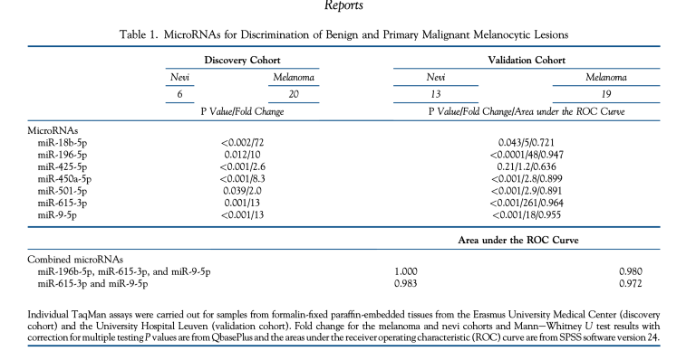
In this study, the authors focused on discriminating miRNA levels in benign versus malignant conjunctival melanocytic lesions and differences in miRNA levels in the conjunctival melanoma with versus without metastases to determine whether it is possible to predict metastatic potential.